Fraunhofer Institute for
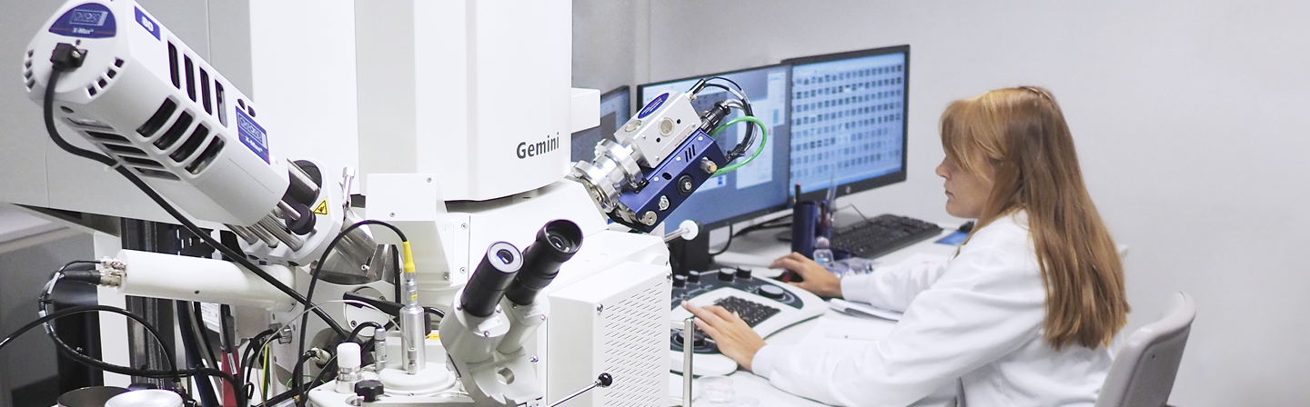
Scanning electron microscopy (SEM)
Analyte
Solid systems; systems with liquid phase
Composites, materials, organic and inorganic substances, thin layers
Equipment
- Device: GeminiSEM 300 (Zeiss), accelerating voltage 0,02 kV ... 30 kV
- Resolution: 0,8 nm at 15 kV
- Magnification: 12x - 2.000.000x
- Everhart-Thornley SE detector
- inLens detector
- ESB detector (energy-selective detector for back-scattering electrons)
- VP mode (low vacuum: 5 - 500 PA)
- Cryo transfer system Alto 2500
- EDX (energy dispersive x-ray microanalysis), Oxford Instruments X-Max 80 (Silcon Drift Detector)
- Digital image capture and archiving
Evaluation methods and targets
- Image analysis by image processing software (Olympus GmbH, Germany): determination of particle sizes and their distribution
- Morphology of fractures, cuts and surfaces with a resolution up to 10 nm
- Qualitative and quantitative elemental analysis
- Identification of morphological structures in materials from natural and synthetic polymers
- Investigation of structure-property-correlations depending on production and growth parameters, respectively, particularly natural polymers
- Investigation of liquid (particularly water-based) systems with cryo-SEM
- Determination of element composition of microstructures
Application examples
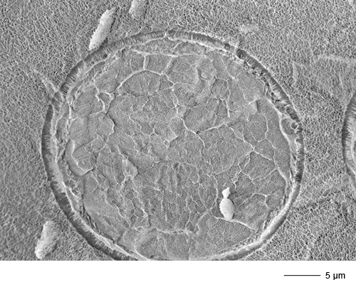
- Humid carbamate fibers: fracture morphology in cryo transfer technology (→ Image gallery)
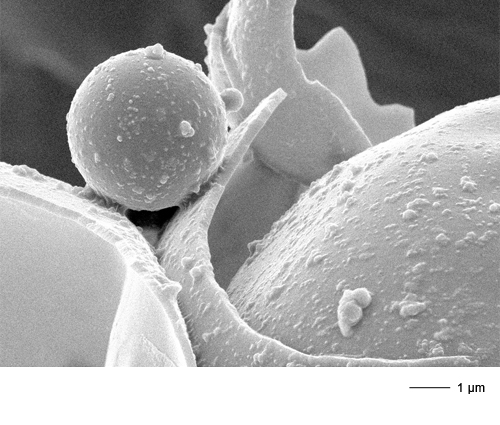
- Microcapsules: surface and wall thickness (→ Image gallery)
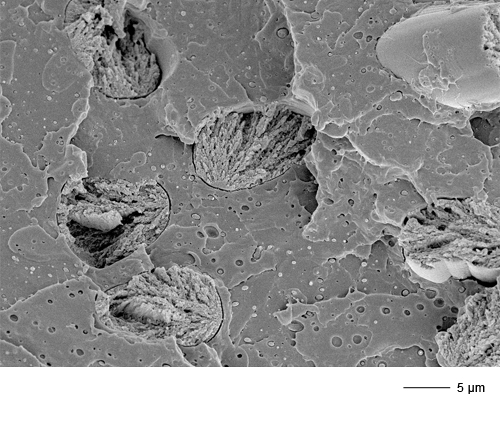
- Composite: PLA-matrix with cellulose fibers, fracture morphology (→ Image gallery)
- Cryo-SEM of formation of pores and crystalline phases in cementitious binder systems