Fraunhofer Institute for
 Optical microscopy
Analyte
Solid and liquid systems;
Composites, materials, organic and inorganic substances, thin layers
Equipment
- Device: polarisation microscope JenLab/pol (Carl Zeiss Jena, Germany), Retarmet2 (Carl Zeiss Jena, Germany)
- Preparation: semi thin cross section with Ultracut S, thin sections with rotationmicrotom VT100E ( Leica, Germany)
- Heating stage (Likam)
- Small angle light scattering
Evaluation methods and targets
- Picture analysis by image processing software (Olympus GmbH, Germany): determination of particle sizes and fiber length, as well its distribution
- Morphology of cuts, (cryo-)fractures and surfaces
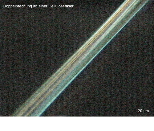
- Determination of birefringence of fibers and layers
- Identification of morphological structures in materials from natural and synthetic polymers
- Investigation of structure-property-correlations depending on production and growth parameters, particularly natural polymers
- Kinetics of crystallization and growth of spherulites
Application examples
- Birefringence at a cellulose fiber (→ Image gallery)
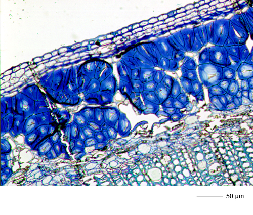
- Semi thin section of a bast fiber (→ Image gallery)
- Growth of spherulites in PLA-fiber-composite
Comparative and supplementary methods
- Transmission electron microscopy (TEM)
- Scanning electron microscopy (SEM)
- ESCA